Bitters: Balancing Agents for the Gut, and Support for Liver/Kidney Detoxification
You can order Dr. Shade’s Bitters No. 9 here.
Bitters: Balancing Agents for the Gut, and Support for Liver/Kidney Detoxification
Bitters: Balancing Agents for the Gut, and Support for Liver/Kidney Detoxification
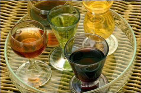
Herbal bitters have a long history of use, and are commonly used in cocktails known as aperitifs and digestifs which are served before and after meals to stimulate appetite and digestion. However, bitter herbs act far beyond the digestive system, and broadly impact the liver, kidneys, skin, immune system, and detoxification pathways.
Bitter herbs broadly encompass many botanicals, and are most often thought of as herbs with effects on the digestive system. Herbal bitters have a long history of use, and are commonly served in cocktails known as aperitifs and digestifs before or after meals to stimulate appetite and digestion. Strong bitter herbs like gentian stimulate stomach acid and other accompanying digestive secretions, while slightly milder bitters such as sweet orange essential oil also calms digestive upset and balances the central nervous system. Bitter herbs act far beyond the digestive system however, and broadly impact the liver, kidneys, skin, immune system, and detoxification pathways.[1] Bitter taste receptors exist not only on the back of the tongue but also throughout the digestive system and beyond.[2] They have been shown to trigger a wide variety of biological processes including regulation of blood sugar and activation of the immune system in response to infections.[3],[4],[5]
The impact of liver, kidney, and gastrointestinal function on detoxification
Although the process of detoxification occurs in every cell of the body, organs in which detoxification occurs at a higher level are the liver and kidneys. It is well known that in individuals with compromised liver or kidney function, dosages of many medications must be altered. The small intestine mucosa also plays an important role in detoxification, as proteins important for all phases of detoxification are expressed at a high level here.[6],[7] When any of these organs (liver, kidneys, intestines) experience less than optimal function, a backup in processing of toxins will occur. With understanding of this, it is easy to see the importance of an integrated process where the intestines, liver, and kidneys are all functioning in an optimal fashion simultaneously.
Gastrointestinal inflammation also effects detoxification, increasing susceptibility of the organism to toxicity from external and internal agents.[8] Leaky gut and damage to the gastrointestinal barrier allows endotoxin, also known as lipopolysaccharide (LPS), to be released into circulation. Exposure to endotoxin effects the other stages of detoxification, downregulating expression of some of the drug and toxin-metabolizing enzymes and Phase III transporters.[9],[10]
The broad impact of the bitter herbs in Dr. Shade’s Bitters No. 9
The proprietary bitters combination of Dr. Shade’s Bitters No. 9 includes dandelion, milk thistle, solidago (goldenrod), gentian, burdock, and essential oils of sweet orange, myrrh, juniper, and clove, which are delivered along with phospholipids that comprise the liposomes in which these herbs are carried. The combination of these herbs is thoughtfully selected to support the gastrointestinal tract, liver, and kidneys in their necessary functions for health and detoxification. Just a small snippet of the vast amounts of research on these herbs is highlighted below.
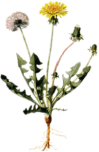
Dandelion
Dandelion is known for its action on the liver and gallbladder, but also acts as an antioxidant and anti-inflammatory, and may have cholesterol lowering effects.[11],[12],[13] In animal models, supplementation with dandelion leaf extract has been shown to alleviate hepatic inflammation associated with a high-fat diet, and protect the liver from alcohol-induced oxidative stress.[14],[15] In the setting of alcohol injury, supplementation with dandelion root extract was observed to increase hepatic antioxidant activity, including glutathione (GSH), GSH-S-transferase, GSH reductase, and GSH peroxidase.
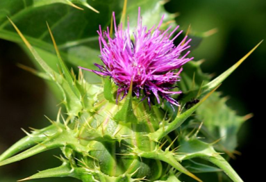
Milk thistle
Milk thistle has been vastly studied for its antioxidative, anti-inflammatory, and hepatoprotective effects in settings including acetaminophen, radiation, iron overload, alcohol and Amanita phalloides (also known as “death cap” mushroom) induced liver damage.[16] Silymarin is the active complex extracted from the seeds of the plant, with the flavonolignan silybin being the most biologically active moiety comprising 50% to 70% of silymarin. Silybin has been observed to inhibit human intestinal β-glucuronidase, blocking the release and
reabsorption of free xenobiotics and their metabolites from their glucuronide conjugates.[17] Silymarin acts as an antioxidant, enhancing hepatic and intestinal GSH levels, and stabilizing membranes by inhibiting membrane peroxidation.[18],[19]
Solidago
Solidago, commonly known as goldenrod, is known for its action on the urinary tract, and is classically used for infections, inflammation, and prevention of kidney stones.[20] Solidago is rich in compounds including flavonoids, phenolic acids, sesquiterpenes, diterpenes, saponins, and several caffeoylquinic acids.[21],[22] Solidago acts as an anti-inflammatory, antimicrobial, diuretic, antispasmodic, and analgesic due to these many compounds found within it.[23] Research has also shown that the flavonoids from solidago have an activating effect on GSH-S-transferases, a critical enzyme in phase II detoxification, in a dose-dependent fashion.[24]
Gentian
Gentian is one of the strongest herbal bitters most often utilized in digestive bitter formulations. Gentian is a digestive toner and modulator of stomach acid secretion, improving function in a state of deficiency but also having a protective effect on conditions such as gastritis or gastric ulcers possibly through prostaglandin pathways.[25] Gentian also acts beyond the digestive system, as the compounds in it exhibit hypoglycemic, hepatoprotective, anti-inflammatory, antioxidant, antimicrobial, immunomodulatory, and adaptogenic properties.[26],[27] As a liver protective agent, gentian has been observed to increase levels of reduced GSH, catalase, superoxide dismutase and GSH peroxidase in various settings of toxin-induced oxidative damage.[28],[29],[30]
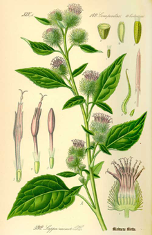
Burdock
Burdock root is commonly utilized in digestive and metabolic balancing formulas, with hypoglycemic, antioxidant, anti-inflammatory, hepatoprotective, and antimicrobial actions.[31] Burdock root has been observed to reverse decreases in GSH and increases in malondialdehyde (a marker of oxidative stress) induced by toxin exposure.[32] Caffeoylquinic acid derivatives from burdock root have been observed to have a strong antioxidant effect, greater than that of α-tocopherol.[33] Burdock extract has been shown to inhibit lipoprotein oxidation while increasing GSH, GSH reductase, GSH peroxidase, GSH-S-transferase and catalase levels.[34]
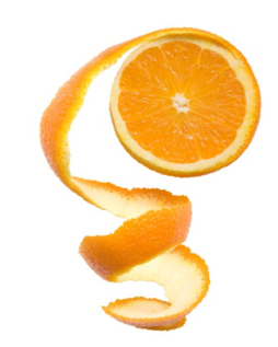
Essential oils of sweet orange, myrrh, juniper, and clove
Each of these oils contains many active compounds, a sense of which we only begin to have from the aromatic expression of its essence. Sweet orange essential oil is derived from the outer peel of the orange, which anyone who has tasted is familiar with its bitter nature. Sweet orange essential oil has been observed to have antibacterial, antifungal, and antioxidant effects.[35],[36] Myrrh has a long history of medicinal use, and is perhaps most recognized for its antimicrobial effects.[37],[38] Additionally, myrrh has been used as an anesthetic, anti-inflammatory, antioxidant, and cholesterol lowering agent.[39]
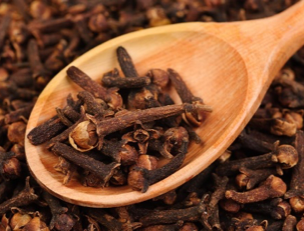
Juniper essential oil also has been shown to have broad antimicrobial action, with traditional use as an antiseptic, antidiarrheal, anti-inflammatory, and astringent, with an affinity for the urinary tract. [40],[41] Juniper also acts as an antioxidant, with metal chelating, free radical, superoxide anion radical, and hydrogen per
oxide scavenging activities.[42],[43] Finally, clove essential oil also has antimicrobial, antiviral, antiulcer, anti-inflammatory and antioxidant properties.[44],[45] As an antioxidant it has a
significant inhibitory effect against hydroxyl radicals and forms complexes with reduced metals.[46],[47],[48]

Author, Dr. Carrie Decker
Dr. Decker is a certified Naturopathic Doctor, graduating with honors from the National College of Natural Medicine (now the National University of Natural Medicine) in Portland, Oregon. Dr. Decker also has graduate degrees in biomedical and mechanical engineering from the University of Wisconsin-Madison and University of Illinois at Urbana-Champaign respectfully. Dr. Decker sees patients at her office in Portland, OR, as well as remotely, with a focus on gastrointestinal disease, mood imbalances, eating disorders, autoimmune disease, chronic fatigue, and skin conditions. Dr. Decker also supports integrative medicine education as a writer and a contributor to various resources.
[1] Shaik FA, et al. Bitter taste receptors: Extraoral roles in pathophysiology. Int J Biochem Cell Biol. 2016 Aug;77(Pt B):197-204. View Abstract
[2] Jaggupilli A, et al. Analysis of the expression of human bitter taste receptors in extraoral tissues. Mol Cell Biochem. 2017 Feb;426(1-2):137-147. View Abstract
[3] Yu Y, et al. Berberine induces GLP-1 secretion through activation of bitter taste receptor pathways. Biochem Pharmacol. 2015 Sep 15;97(2):173-7. View Abstract
[4] Lee RJ, Cohen NA. The emerging role of the bitter taste receptor T2R38 in upper respiratory infection and chronic rhinosinusitis. Am J Rhinol Allergy. 2013 Jul-Aug;27(4):283-6. View Abstract
[5] Gaida MM, et al. Sensing developing biofilms: the bitter receptor T2R38 on myeloid cells. Pathog Dis. 2016 Apr;74(3). View Full Paper
[6] Doherty MM, Charman WN. The mucosa of the small intestine: how clinically relevant as an organ of drug metabolism? Clin Pharmacokinet. 2002;41(4):235-53. View Abstract
[7] Berggren S, et al. Gene and protein expression of P-glycoprotein, MRP1, MRP2, and CYP3A4 in the small and large human intestine. Mol Pharm. 2007 Mar-Apr;4(2):252-7. View Abstract
[8] Ganey PE, Roth RA. Concurrent inflammation as a determinant of susceptibility to toxicity from xenobiotic agents. Toxicology. 2001 Dec 28;169(3):195-208. View Abstract
[9] Tang W, et al. Endotoxin downregulates hepatic expression of P-glycoprotein and MRP2 in 2-acetylaminofluorene-treated rats. Mol Cell Biol Res Commun. 2000 Aug;4(2):90-7. View Abstract
[10] Kalitsky-Szirtes J, et al. Suppression of drug-metabolizing enzymes and efflux transporters in the intestine of endotoxin-treated rats. Drug Metab Dispos. 2004 Jan;32(1):20-7. View Full Paper
[11] Schütz K, Carle R, Schieber A. Taraxacum--a review on its phytochemical and pharmacological profile. J Ethnopharmacol. 2006 Oct 11;107(3):313-23. View Abstract
[12] González-Castejón M, et al. Diverse biological activities of dandelion. Nutr Rev. 2012 Sep;70(9):534-47. View Abstract
[13] Choi UK, et al. Hypolipidemic and antioxidant effects of dandelion (Taraxacum officinale) root and leaf on cholesterol-fed rabbits. Int J Mol Sci. 2010 Jan 6;11(1):67-78. View Full Paper
[14] Davaatseren M, et al. Taraxacum official (dandelion) leaf extract alleviates high-fat diet-induced nonalcoholic fatty liver. Food Chem Toxicol. 2013 Aug;58:30-6. View Abstract
[15] You Y, et al. In vitro and in vivo hepatoprotective effects of the aqueous extract from Taraxacum officinale (dandelion) root against alcohol-induced oxidative stress. Food Chem Toxicol. 2010 Jun;48(6):1632-7. View Abstract
[16] Abenavoli L, et al. Milk thistle in liver diseases: past, present, future. Phytother Res. 2010 Oct;24(10):1423-32. View Abstract
[17] Kim DH, et al. Silymarin and its components are inhibitors of beta-glucuronidase. Biol Pharm Bull. 1994 Mar;17(3):443-5. View Abstract
[18] Valenzuela A, et al. Selectivity of silymarin on the increase of the GSH content in different tissues of the rat. Planta Med. 1989 Oct;55(5):420-2. View Abstract
[19] Rui YC. Advances in pharmacological studies of silymarin. Mem Inst Oswaldo Cruz. 1991;86 Suppl 2:79-85. View Full Paper
[20] Melzig MF. [Goldenrod--a classical exponent in the urological phytotherapy]. Wien Med Wochenschr. 2004 Nov;154(21-22):523-7. View Abstract
[21] Bradette-Hébert ME, et al. A new labdane diterpene from the flowers of Solidago canadensis. Chem Pharm Bull (Tokyo). 2008 Jan;56(1):82-4. View Full Paper
[22] Bohlmann F, et al. Sesquiterpene and diterpene derivatives from Solidago species. Phytochemistry. 1980 Jan;19(12):2655-61. View Abstract
[23] Yarnell E. Botanical medicines for the urinary tract. World J Urol. 2002 Nov;20(5):285-93. View Full Paper
[24] Apáti P, et al. In-vitro effect of flavonoids from Solidago canadensis extract on GSH S-transferase. J Pharm Pharmacol. 2006 Feb;58(2):251-6. View Abstract
[25] Niiho Y, et al. Gastroprotective effects of bitter principles isolated from Gentian root and Swertia herb on experimentally-induced gastric lesions in rats. Journal of natural medicines. 2006 Jan;60(1):82-8. View Abstract
[26] Dinda B, et al. Naturally occurring iridoids, secoiridoids and their bioactivity. An updated review, part 3. Chem Pharm Bull (Tokyo). 2009 Aug;57(8):765-96. View Full Paper
[27] Ghisalberti EL. Biological and pharmacological activity of naturally occurring iridoids and secoiridoids. Phytomedicine. 1998 Apr;5(2):147-63. View Abstract
[28] Mihailović V, et al. Hepatoprotective effects of Gentiana asclepiadea L. extracts against carbon tetrachloride induced liver injury in rats. Food Chem Toxicol. 2013 Feb;52:83-90. View Abstract
[29] Lian LH, et al. Gentiana manshurica Kitagawa reverses acute alcohol-induced liver steatosis through blocking sterol regulatory element-binding protein-1 maturation. J Agric Food Chem. 2010 Dec 22;58(24):13013-9. View Abstract
[30] Wang AY, et al. Gentiana manshurica Kitagawa prevents acetaminophen-induced acute hepatic injury in mice via inhibiting JNK/ERK MAPK pathway. World J Gastroenterol. 2010 Jan 21;16(3):384-91. View Abstract
[31] Chan YS, et al. A review of the pharmacological effects of Arctium lappa (burdock). Inflammopharmacology. 2011 Oct;19(5):245-54. View Abstract
[32] Lin SC, et al. Hepatoprotective effects of Arctium lappa on carbon tetrachloride- and acetaminophen-induced liver damage. Am J Chin Med. 2000;28(2):163-73. View Abstract
[33] Maruta Y, Kawabata J, Niki R. Antioxidative caffeoylquinic acid derivatives in the roots of burdock (Arctium lappa L.). Journal of Agricultural and Food Chemistry. 1995 Oct;43(10):2592-5. View Abstract
[34] Wang BS, et al. Protective effects of burdock (Arctium lappa Linne) on oxidation of low-density lipoprotein and oxidative stress in RAW 264.7 macrophages. Food Chemistry. 2007 Dec;101(2):729-38. View Abstract
[35] Geraci A, et al. Essential oil components of orange peels and antimicrobial activity. Nat Prod Res. 2016 Aug 18:1-7. View Abstract
[36] Guimarães R, et al. Targeting excessive free radicals with peels and juices of citrus fruits: grapefruit, lemon, lime and orange. Food Chem Toxicol. 2010 Jan;48(1):99-106. View Abstract
[37] Dolara P, et al. Local anaesthetic, antibacterial and antifungal properties of sesquiterpenes from myrrh. Planta Med. 2000 May;66(4):356-8. View Abstract
[38] Sheir Z, et al. A safe, effective, herbal antischistosomal therapy derived from myrrh. Am J Trop Med Hyg. 2001 Dec;65(6):700-4. View Full Paper
[39] Shen T, et al. The genus Commiphora: a review of its traditional uses, phytochemistry and pharmacology. J Ethnopharmacol. 2012 Jul 13;142(2):319-30. View Abstract
[40] Pepeljnjak S, et al. Antimicrobial activity of juniper berry essential oil (Juniperus communis L., Cupressaceae). Acta Pharm. 2005 Dec;55(4):417-22. View Abstract
[41] Bais S, et al. A Phytopharmacological Review on a Medicinal Plant: Juniperus communis. Int Sch Res Notices. 2014 Nov 11;2014:634723. View Full Paper
[42] Miceli N, et al. Comparative analysis of flavonoid profile, antioxidant and antimicrobial activity of the berries of Juniperus communis L. var. communis and Juniperus communis L. var. saxatilis Pall. from Turkey. J Agric Food Chem. 2009 Aug 12;57(15):6570-7. View Abstract
[43] Elmastaş M, et al. A study on the in vitro antioxidant activity of juniper (Juniperus communis L.) fruit extracts. Analytical letters. 2006 Jan;39(1):47-65. View Abstract
[44] Santin JR, et al. Gastroprotective activity of essential oil of the Syzygium aromaticum and its major component eugenol in different animal models. Naunyn Schmiedebergs Arch Pharmacol. 2011 Feb;383(2):149-58. View Abstract
[45] Chaieb K, et al. The chemical composition and biological activity of clove essential oil, Eugenia caryophyllata (Syzigium aromaticum L. Myrtaceae): a short review. Phytother Res. 2007 Jun;21(6):501-6. View Abstract
[46] Misharina TA, et al. [Antiradical properties of essential oils and extracts from clove bud and pimento]. Prikl Biokhim Mikrobiol. 2015 Jan-Feb;51(1):99-104. View Abstract
[47] Jirovetz L, et al. Chemical composition and antioxidant properties of clove leaf essential oil. J Agric Food Chem. 2006 Aug 23;54(17):6303-7. View Abstract
[48] Ito M, et al. Antioxidant action of eugenol compounds: role of metal ion in the inhibition of lipid peroxidation. Food Chem Toxicol. 2005 Mar;43(3):461-6. View Abstract
WHAT'S UP WITH MERCURY?
"Mercury is the hottest, the coldest, a true healer, a wicked murderer, a precious medicine and a deadly poison, a friend that can flatter and lie." – Woodall from The Surgeon’s Mate, London, 1639
MERCURY
Chemical Element
Mercury is a chemical element with symbol Hg and atomic number 80. It is commonly known as quicksilver and was formerly named hydrargyrum. Wikipedia
Symbol: Hg
Melting point: -37.89°F (-38.83°C)
Electron Configuration: [Xe] 4f14 5d10 6s2
Atomic number: 80
Boiling point: 674.1°F (356.7°C)
Discovered: 2000 BC
Atomic mass: 200.59 ± 0.02 u
The greatest source of mercury in the biosphere is currently of human origin. Mercury is number three on the Agency for Toxic Substances & Disease Registry (ATSDR) 2011 Substance Priority List. Although mercury is a naturally occurring element, most of it is sequestered in subterranean rock formations and coal beds. Two-thirds of the mercury entering the biosphere comes from man-made sources, including industrial plants, coal burning and incinerators; the additional one-third is emitted from natural sources. Many former chlor-alkali facilities (for producing chlorine and sodium hydroxide) are major point sources of mercury to aquatic ecosystems and are currently designated Superfund sites. Mercury is released into the air or directly into water bodies and makes its way into lakes and estuaries, where some of it settles to the bottom. Bacteria living in the mud of lake, river and estuary bottoms helps transform mercury into methylmercury (see below).
But mercury is considered a global pollutant, as it's capable of spreading far beyond its source area. The arctic, for example, has no known sources of mercury, but it harbors mercury-contaminated fish, and recent studies indicate that whales feeding in the arctic have high levels of mercury in their tissue.
What is Methylmercury? Methylmercury is an extremely toxic form of mercury that biomagnifies in aquatic food chains. It is a potent neurotoxin and the easiest form for animals to store in their tissue. It harms the brain, affecting memory, understanding and movement. Studies have shown that mercury exposure in humans can result in developmental delays in children, motor impairment, cardiovascular effects and, in acute cases, death. Its effects have been studied in fish, whales, seals and seabirds. Methylmercury binds to proteins and easily crosses cell membranes, including the blood-brain barrier and the placenta. Affected wildlife, such as loons, develop behaviors that ultimately reduce their chances for survival and reproduction. Studies conducted on human populations have estimated that between 200,000 and 400,000 children in the United States alone are born each year with pre-natal exposure to methylmercury sufficient to put them at risk of neurologic impairment.
Inorganic mercury is the term used to refer to mercuric ion (HgII). Inorganic mercury is highly toxic but not very mobile. Inorganic mercury in sediments, soils and food sources does not pass easily into biological tissues. However, once inside of the tissue, inorganic mercury is very difficult to remove. Inorganic mercury accumulates in tissues when a more mobile form of mercury such as elemental mercury vapor, methylmercury or ethylmercury enters the tissue and breaks down into inorganic mercury. In biological tissues, most organic forms of mercury will eventually break down into inorganic mercury.
Ethylmercury
Like methylmercury, ethylmercury is an organic form of mercury. Ethylmercury can be present in sediments or petroleum hydrocarbons. Ethylmercury is also used as a component in vaccine preservatives (thimerosal). Vaccination is the most common exposure route for this organic form of mercury. Like methylmercury, ethylmercury can move easily into biological tissues. However, ethylmercury tends to break down into inorganic mercury more rapidly than methylmercury.
Bring the Power of Intravenous Therapy into a Convenient Oral Liposomal Delivery
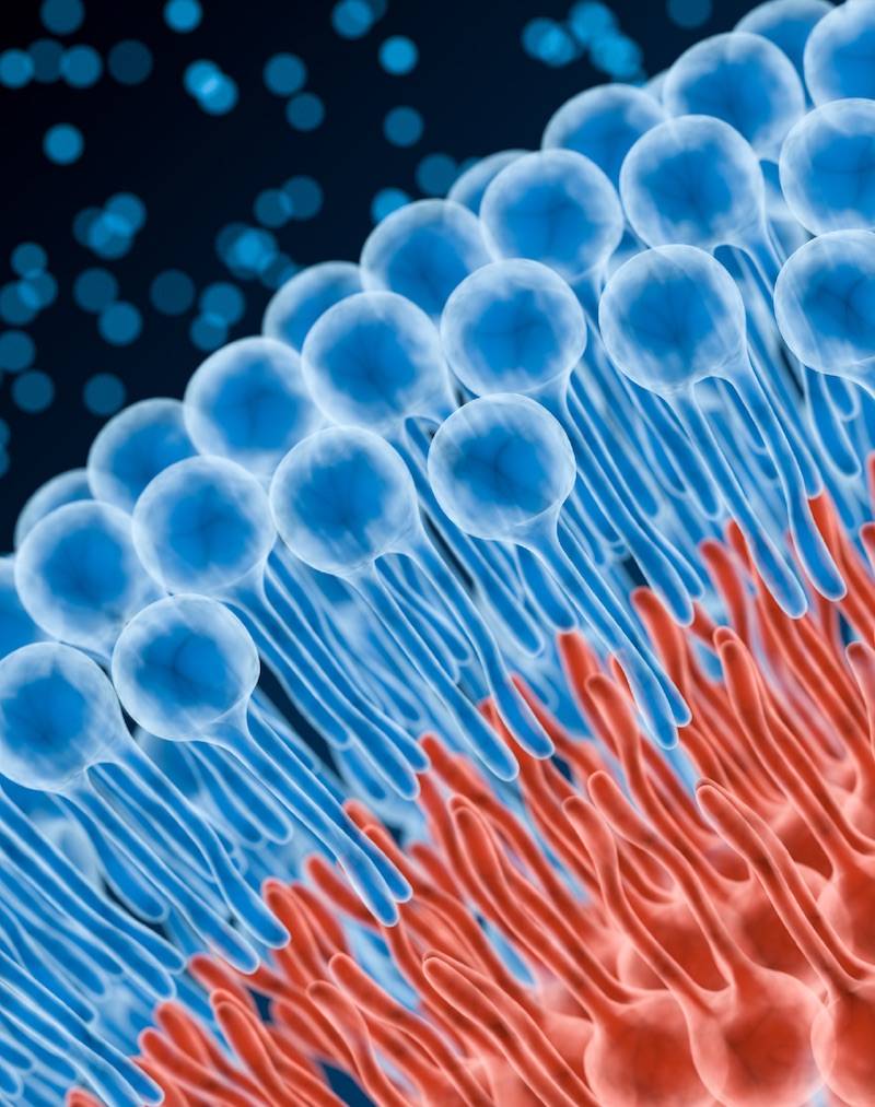
Liposomes are microscopic single- to multi-layer spheres made of phospholipids (the basic building blocks of cell membranes). Phospholipids encapsulate a compound, such as glutathione, in order to bypass the digestive processes that normally degrade or limit the compounds' absorption. Liposomes demonstrate the ability to cross the blood-brain barrier, deposit their cargo intracellularly, and enhance lymphatic circulation of therapeutic compounds. Additionally, the phospholipids which compose the liposome fuse with and feed the cell membranes. Providing phospholipids in this way ensures proper membrane function to facilitate the absorption of nutrients and the excretion of cellular waste products and toxins. In fact, phospholipid therapy, using both injectable forms and oral forms, has a long and solid clinical history for repair and maintenance of liver function, circulatory health, and neurological health.

At their highest expression, liposomes
bring the power of intravenous therapy into a convenient oral delivery.
However, all liposomes are not created equally. Most liposomes on the
market use low grades of phospholipids (e.g. raw lecithin) instead of
the high-phosphatidylcholine phospholipid mixes necessary for a good
liposome. Nutraceutical manufacturers also typically use cheap shear
methods resulting in large (200–600nm), poorly-absorbed particles.
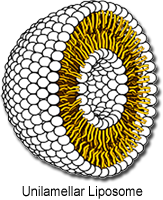
Quicksilver’s Etheric Delivery™ Phospholipid Encapsulation System improves upon basic liposomal Delivery technology
with smaller, more stable, tightly distributed single-layer spheres
(called unilamellar vessicles) made from the highest-grade ingredients
available. These small vessicles begin absorbing as soon as
they hit your mouth so you can get high absorption before they are
altered by the harsh processes in the intestines. Quicksilver Scientific
uses the same high-tech equipments, rigorous processes, and tight
particle size controls used by pharmaceutical companies. Don't settle
for the low grade products now flooding the market, and certainly don't
fall for the ploy that you can make your own "bathtub gin" with a
blender and a sonic jewelery cleaner! Look for the clear difference of
Quicksilver Scientific's Etheric Delivery™ products. Our liposomal
vitamins (like the totally unique liposomal B-Complex) are superior.
Period.